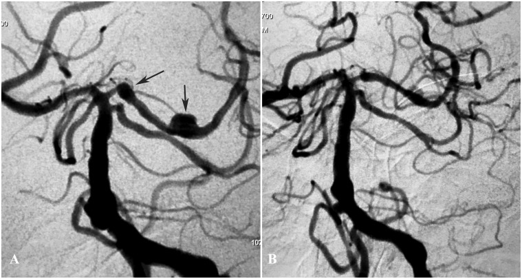
Figure 1.
(a) Left vertebral angiogram showing the left posterior cerebral artery two small fusiform aneurysms (arrows). (b) Immediate postoperative view showing the single Pipeline flow-diverting stent (2.75 × 20 mm) placed in the left posterior cerebral artery, resulting in contrast stasis within the sac.

