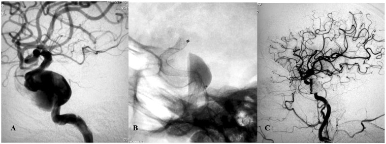
Figure 4.
(a) Lateral angiogram showing a giant cavernous internal carotid artery aneurysm. (b) Intraoperative view showing the Pipeline flow-diverting stent (3.75 × 30 mm) opening to the normal size of the parent artery. Note the contrast stagnation within the sac. (c) Angiography after placement of coils showing complete occlusion of the aneurysm and reconstruction of the parent artery.

