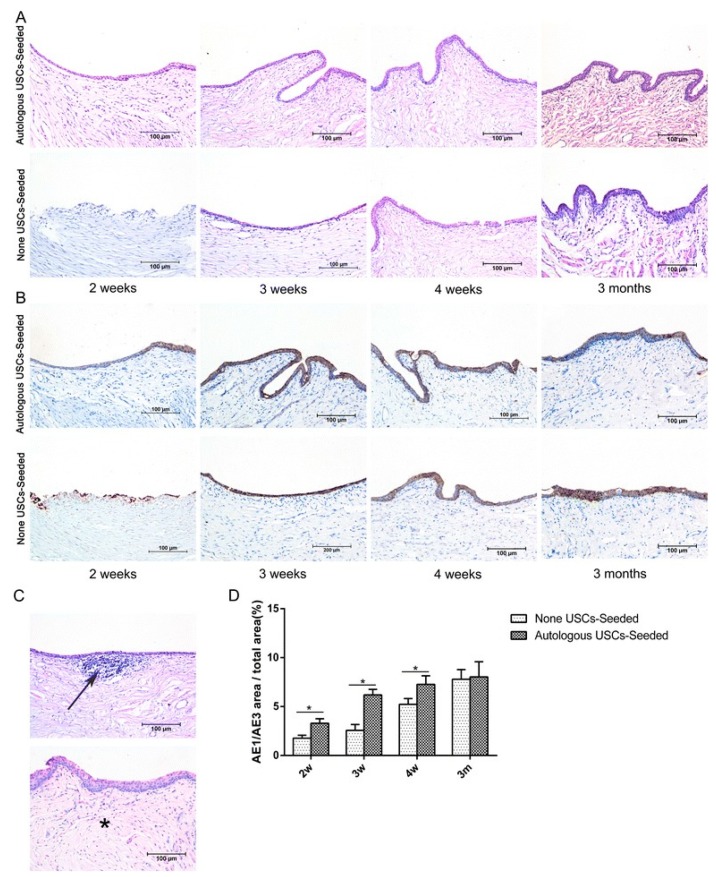
Figure 2.
Histopathological evaluation of urothelium regeneration. H&E (A) and AE1/AE3 IHC (B). The USC-seeded 5% PAA-treated SIS group showed better multilayered urothelial tissue over the different time frames in comparison to 5% PAA-treated SIS only group. (C) Infiltration of inflammatory cells (arrow) and fibrosis (*) were observed in the 5% PAA-treated SIS only group. Scale bar = 200 μm. (D) Image analysis of the AE1/AE3-positive area to the total area at each time point in the two groups. *Statistically significant (p < 0.05). m: months, USC: urine-derived stem cell, w: weeks. Reproduced with permission from [68].

