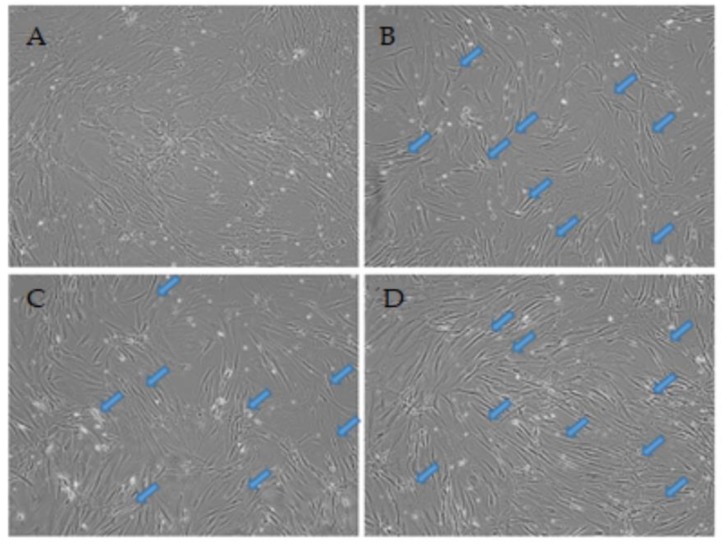
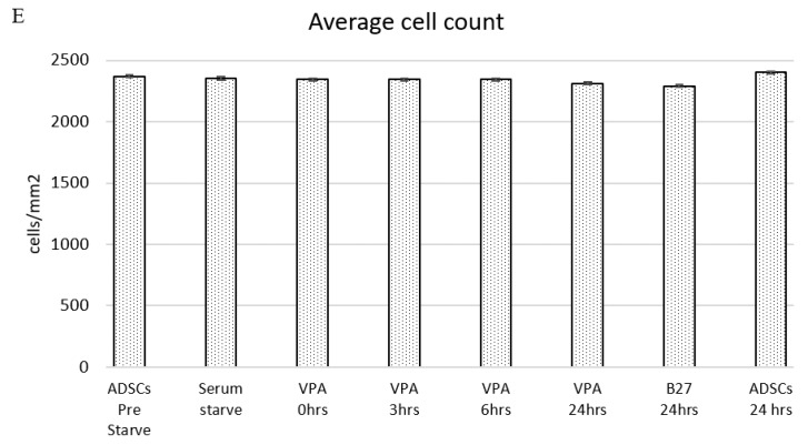
Figure 1.
Live cell images of the temporal differentiation of human adipose derived stem cells (ADSCs) induced with 0.2 mM valproic acid (VPA) at (A) 0 h, (B) 3 h, (C) 6 h, and (D) 24 h at 10× magnification. The cellular morphology changes rapidly through the time points, with cells adopting a more slender and bipolar orientation with neurite extensions (arrows). (E) Average cell count across the treatments including all controls with standard error bars. Relatively minimal changes in numbers are shown across all treatments. A Student’s t-test revealed no significant change in cell numbers.


