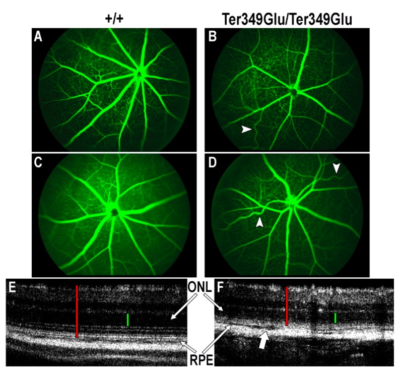
Figure 2.
Ter349Glu rhodopsin knock-in mouse retina exhibits both vascular and laminar abnormalities. (A–D) Utilizing fluorescein angiography (FA), the state of the retinal vasculature of 4-week-old +/+ (A,B) and Ter349Glu/Ter349Glu (C,D) mice was examined. Abnormal phenotypes varied in severity among mice, with overall attenuated retinal vessels and tortuous retinal vessels (arrowheads) being commonplace amongst all mice examined. (E,F) Optical coherence tomography (OCT) was used to examine the retinas of 4-week-old +/+ (E) and Ter349Glu/Ter349Glu mice (F) for architectural abnormalities. Ter349Glu/Ter349Glu mice exhibited thinning of the outer nuclear layer (ONL) and patches of varying degrees of separation among the choroid, retinal pigment epithelium (RPE), and photoreceptors (block arrow), indicative of edema. Retinal thickness (red calipers) = 240 μm (E) and 180 μm (F); ONL (green calipers) = 60 μm (E) and 50 μm (F).

