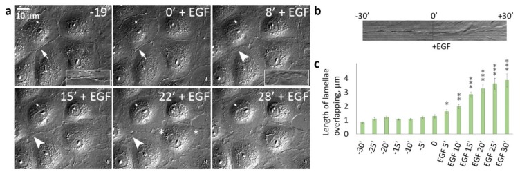
Figure 2.
Disappearance of contact paralysis in IAR-20 epithelial cells after the addition of EGF. (a) Selected frames from Video S2. The cell-cell boundaries (indicated with arrows and arrowheads) are enlarged in boxed regions. In control cells, the thin “scars” of cell-cell contacts are seen at the cell-cell boundaries (arrows). In the presence of EGF, cell-cell interfaces became highly unstable (arrowheads) and overlapping lamellae were seen at the cell-cell boundaries. Asterisks indicate sites of disruption of cell-cell contacts. (b) Kymograph generated at the site of the cell-cell contact shows contact paralysis before EGF addition and appearance of protrusive activity caused by addition of EGF. (c) Length of lamellae overlapping at the cell-cell boundaries. * p < 0.05, ** p < 0.001, *** p < 0.0001.

