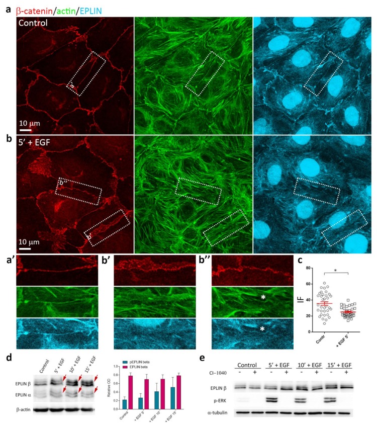
Figure 8.
Effects of EGF on EPLIN in IAR-20 cells. (a), (a’) In control IAR-20 cells, EPLIN colocalizes with the circumferential actin bundles at cell-cell boundaries. (b) Addition of EGF leads to release of EPLIN from the zones of disorganization or disappearance of the circumferential bundles (Figure 8b’,b’’). EPLIN colocalizes with the remaining intact circumferential bundle (Figure 8b’’, asterisk). (c) EPLIN fluorescence intensity at the cell-cell boundaries in control and EGF-treated cells. Circles and squares represent individual cells, N = 35, * p < 0.001. (d) Western blot analysis of EPLIN phosphorylation (5% PAAG). Arrows indicate up-shifted bands of phosphorylated EPLIN in the cells treated with EGF. β-actin was used as loading control. Densitometry results are averaged across three independent experiments. Data are presented as mean ± SEM. (e) MEK inhibitor CI-1040 (4 µM), which inhibits phosphorylation of ERK (p-ERK), significantly decreases the levels of phosphorylated EPLIN at 10 min and 15 min after the addition of EGF. α-tubulin was used as a loading control.

