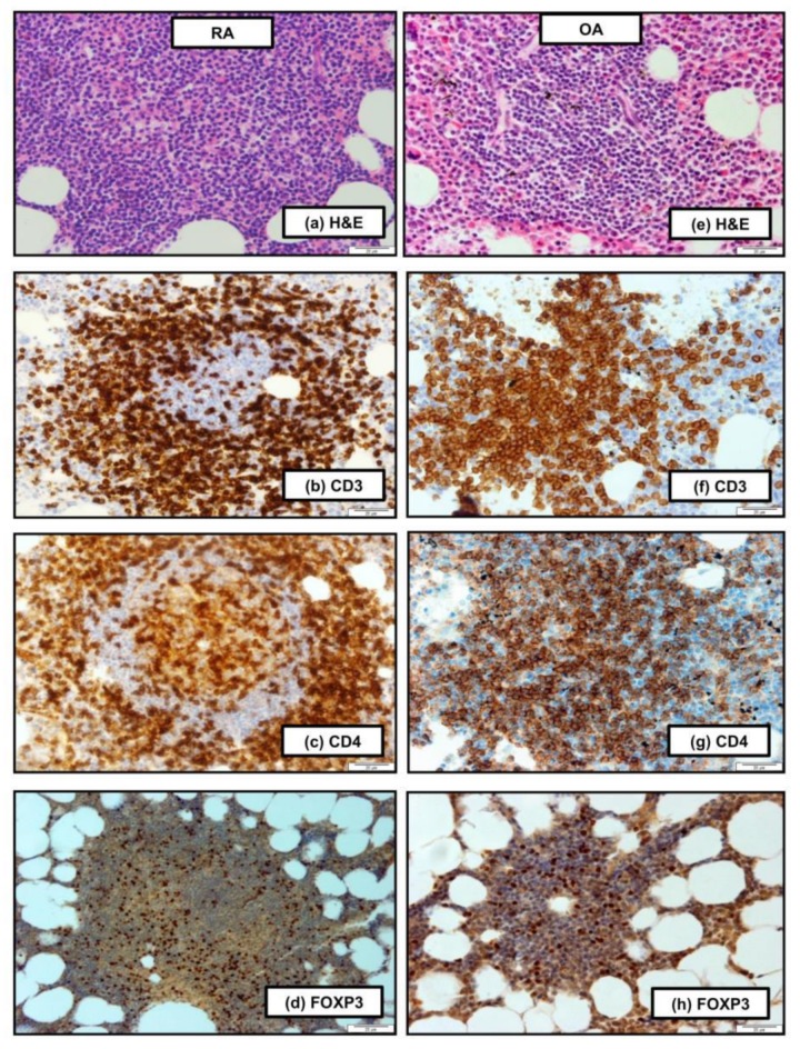
Figure 1.
Histopathological features of the bone marrow (BM) of patients with rheumatoid arthritis (RA) (a–d) and osteoarthritis (OA) (e–h). (a) Nodular lymphocytic infiltration with germinal center formation (hematoxylin and eosin [H&E] stain, 100×). (b) CD3+ T cells in the marginal and mantle zone. (c) CD4+ T cells in the lymphoid follicle. (d) Nuclear expression of FOXP3 in cells localized in the lymphoid follicle. (b–d: EnVision stain, 100×). (e) H&E staining shows visible nodular lymphocytic infiltration, 100×. (f,g) Most of the lymphocytes in the lymphoid follicle revealed CD3 and CD4 expression. (h) FOXP3 in nuclear localization in cells of the lymphoid follicle (f–h: EnVision stain, 100−). Scale bar, 20 μm. Histology staining was done on five patients in each group while one representative is shown.

