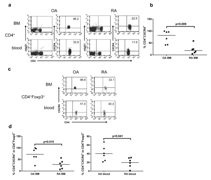
Figure 3.
Expression of CXCR4 by CD4+ and CD4+FOXP3+ cells from the bone marrow and peripheral blood of OA and RA patients. (a) Representative dot plots show CXCR4 expression by gated CD4+ cells in the BM and peripheral blood of the same patient. (b) CXCR4 expression by CD4+ T cells in OA and RA BM. (c) Representative dot plots show CXCR4 expression by gated CD4+FOXP3+ regulatory T cells (Tregs) in the BM and peripheral blood of the same patient. (d) CXCR4 expression by gated CD4+FOXP3+ Tregs in the BM (chart on the left) and peripheral blood (chart on the right) of OA and RA patients. In all the cases, differences between groups of patients were analyzed by Mann–Whitney U-test. Numbers depicted on dot plots show the frequencies of subset expressing CXCR4. Individual results are shown as dots and median as a bar on charts (n = 6 subjects per group). OA/RA BM/blood—cells isolated from the BM/peripheral blood of patients with OA/RA, respectively.

