Figure 1.
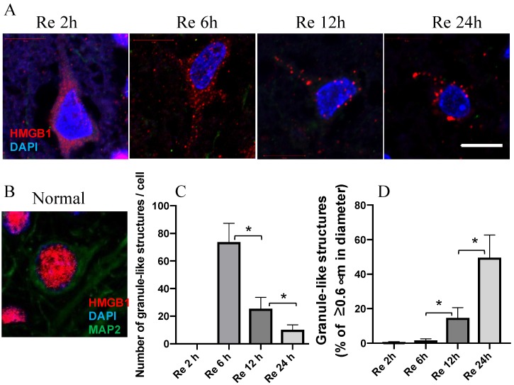
HMGB1 translocation at the indicated time intervals. (A) HMGB1 staining in a typical neuron in the parietal cortex at 2, 6, 12, and 24 h after reperfusion of MCAO. (B) HMGB1 staining in a typical neuron of cortex in normal rat brain. (C) The number of granule-like structures with the diameter more than 0.3 μm per cell in the cytosol at the indicated time intervals after reperfusion of MCAO. (D) The ratios of the number of granule-like structures with more than 0.6 μm in diameter vs. total number of granule-like structures in the cytosol were determined at the indicated time intervals after reperfusion of MCAO. Scale bar: 10 μm. (n = 6, * p < 0.05)

