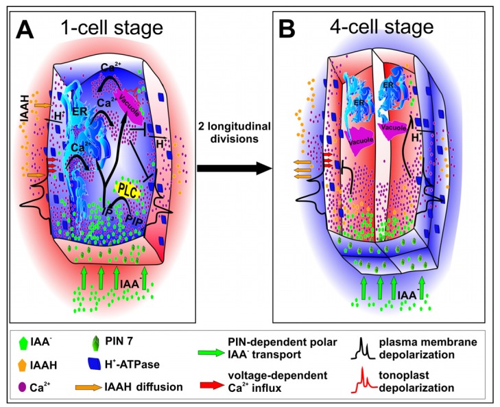
Figure 2.
A hypothetical model of calcium ion release. (A) Transport of auxin ions to the apical cell is provided by polar localization of PIN7 proteins. The auxin influx induces plasma membrane depolarization, followed by an opening of calcium channels. Auxin and calcium ions regulate the activity of phospholipase C (PLC) which catalyzes conversion of PIP2 to IP3. IP3 induces opening ligand-gated calcium channels by binding to specific receptors in ER or vacuole. An elevated concentration of calcium ions activates also voltage-dependent channels in a vacuole. On the other hand, high concentration of calcium ions triggers inhibition of plasma membrane H+-ATPase which results in apoplast alkalization. High pH of an apoplast reduces auxin-induced plasma membrane depolarization and may act as self-attenuating mechanism of auxin impact. (B) After two longitudinal divisions the cell volume is 4-fold smaller but the surface of vertical membranes of daughter cells is only halved. All this results in faster achievement of high calcium concentration and following inhibition of plasma membrane H+-ATPase. Apoplast alkalization reduces auxin-induced depolarization of plasma membrane. In turn, cytosol acidification results in auxin protonation which allows for its diffusion from cytoplasm. Thus, both auxin concentration and calcium ion release from ER and vacuole are diminished.

