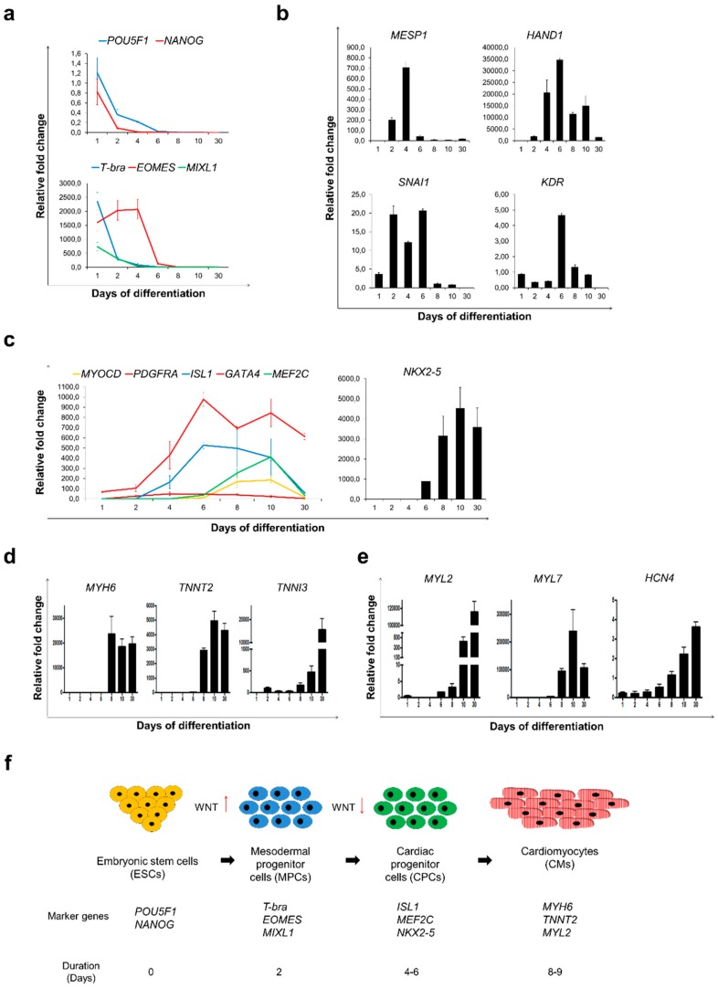
Figure 2.
Sulindac treatment was sufficient to accelerate cardiac differentiation and maturation of cardiomyocytes. Gene expression analysis by qRT-PCR was performed during Sulindac-based cardiac differentiation protocol for; (a) markers for pluripotency: POU5F1 and NANOG (Top) and early mesoderm: T-bra, EOMES and MIXL1 (bottom), (b) markers for cardiac mesoderm: MESP1, HAND1, SNAI1 and KDR, (c) markers for cardiac progenitor cells: MYOCD, PDGFRA, ISL1, GATA4, MEF2C and NKX2-5, (d) Cardiac-specific genes MYH6, TNNT2 and TNNI3, (e) ventricular, atrial and nodal sub-type-specific markers MYL2, MYL7 and HCN4. Samples collected for this analysis were photographed and presented in Figure 1d. All Ct values are normalised using GAPDH and relative fold change was calculated using Day 0 HES3 cells as control. Error bars, ±SEM; n = 3 independent biological replicates; (f) Schematic representation of transition of HES3 cells from pluripotent stem cells to cardiomyocytes represented by stage-specific expressed marker genes.

