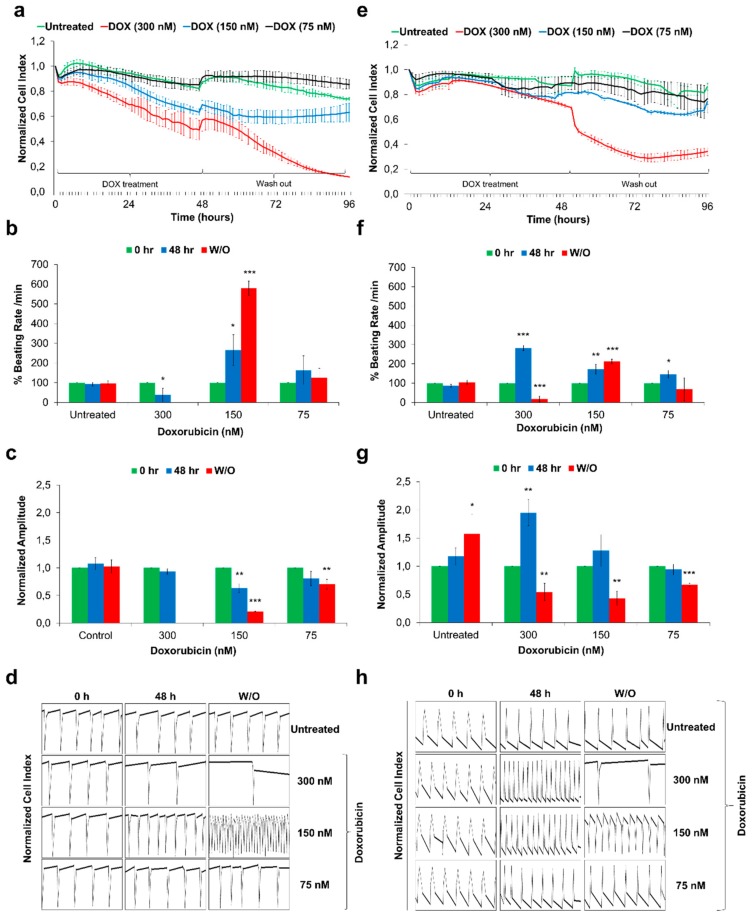
Figure 4.
Functional studies of Doxorubicin-exposed Sulindac-derived HES3-CMs and hiPSC-CMs using the xCELLigence RTCA Cardio system. The HES3-CMs and hiPSC-CMs were cultured in fibronectin-coated E-cardio plates and treated with Doxorubicin for 48 h followed by 48 h drug washout (W/O). Cells were monitored using xCELLIgence RTCA Cardio system for real time changes; (a,e) The representative graphs displays DOXO exposure induces cytotoxicity and drop in Cell Index values in HES3-CMs and hiPSC-CMs respectively. Error bars, ±SEM; n = 3 independent biological replicates; (b,f) The representative graphs displays DOXO exposure induces changes in % beating rates in HES3-CMs and hiPSC-CMs respectively. Error bars, ±SEM; n = 3 independent biological replicates; (Student’s t test, * p ≤ 0.05, ** p ≤ 0.01, *** p ≤ 0.001) (c,g) The representative graphs displays DOXO exposure induces changes in beating amplitude in HES3-CMs and hiPSC-CMs respectively. Error bars, ±SEM; n = 3 independent biological replicates; (Student’s t test, * p ≤ 0.05, ** p ≤ 0.01, *** p ≤ 0.001) (d,h) Representative 12 s beating traces of HES3-CMs and hiPSC-CMs after DOXO exposures and during drug washout. Y-axis represents Normalised Cell Index. Beating activity demonstrates the development of arrhythmic beating upon DOXO exposure. Error bars, ±SEM; n = 3 independent biological replicates.

