Figure 7.
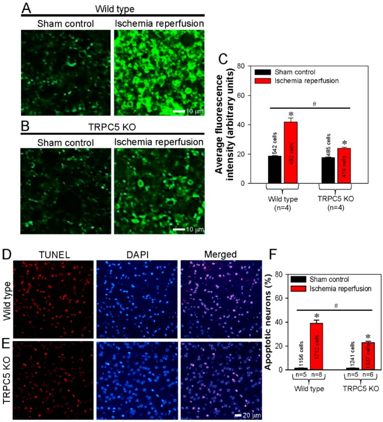
Knockout of TRPC5 protects cerebral neurons from ischemia-reperfusion-induced apoptosis. (A, B) Representative images and summarized data showing phosphatidylserine (PS) externalization as determined using an annexin V-FITC assay in neurons of cerebral slices obtained from wild-type (A, C) and TRPC5 knockout (TRPC5 KO) (B, C) mice subjected to sham surgery (sham control) or cerebral ischemia-reperfusion. (C) Summary of average fluorescence intensity (arbitrary units) of PS externalized neurons in cerebral slices obtained from wild-type and TRPC5 KO mice subjected to sham surgery (control) or cerebral ischemia-reperfusion. (D, E) Following surgery, TUNEL assays were performed to visualize apoptotic mouse cerebral neurons as red fluorescence signals in the neurons of cerebral slices obtained from wild-type (D) or TRPC5 KO (E) mice subjected to cerebral ischemia-reperfusion. Cell nuclei were stained blue with DAPI. Merged images show apoptotic signals overlapping with cell nuclei. (F) Summary of the percentages of apoptotic neurons in cerebral slices obtained from wild-type and TRPC5 KO mice subjected to sham surgery (control) or cerebral ischemia–reperfusion. Values represent the mean ± SEM (n = 5-8); *p < 0.05, control vs. ischemia-reperfusion with a two-tailed unpaired Student’s t test; #p < 0.05, wild-type ischemia-reperfusion vs. TRPC5 KO ischemia–reperfusion with two-way ANOVA followed by the Tukey’s post-hoc comparison test.

