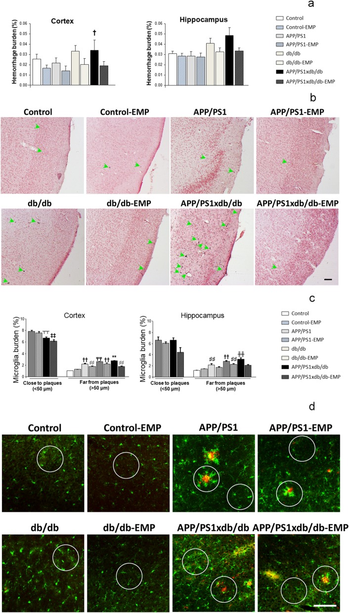
Fig. 4.
Spontaneous bleeding and inflammation were reduced after EMP treatment. a EMP reduced hemorrhage burden in the cortex, and while a similar profile was observed in the hippocampus, differences were not statistically significant (cortex [F(7, 262) = 2.65, †p = 0.011 vs. control, control-EMP, APP/PS1, and APP/PS1-EMP]; hippocampus [F(7, 118) = 01.85, p = 0.081]) (control n = 8, control-EMP n = 6, APP/PS1 n = 5, APP/PS1-EMP n = 4, db/db n = 7, db/db-EMP n = 6, APP/PS1xdb/db n = 4, APP/PS1xdb/db-EMP n = 6). b Illustrative example of Prussian blue staining in all groups under study. Green arrows point at individual hemorrhages. Scale bar = 100 μm. c Cortical microglia burden was lower in the proximity of SP in APP/PS1xdb/db mice ([F(3, 534) = 7.036], ‡‡p < 0.01 vs. APP/PS1 and APP/PS1-EMP, ┬┬p < 0.01 vs. APP/PS1]), while in SP-free areas, increased burden was reduced after EMP treatment ([F(7, 4465)=137.36, **p < 0.01 vs. the rest of the groups, ₸₸p < 0.01 vs. control, control-EMP, APP/PS1, APP/PS1-EMP, db/db-EMP, and APP/PS1xdb/db-EMP; ††p < 0.01 vs. control, control-EMP, APP/PS1-EMP, and APP/PS1xdb/db-EMP, ##p < 0.01 vs. APP/PS1-EMP and APP/PS1xdb/db-EMP]). A similar profile was observed in the hippocampus, and microglia burden was reduced after EMP treatment in SP-free areas (close to plaques ([F(3, 39)=2.21, p = 0.91]); far from plaques [F(7, 768) = 23.79, ╫╫p < 0.01 vs. control, control-EMP, APP/PS1, APP/PS1-EMP, and APP/PS1xdb/db-EMP, ††p < 0.01 vs. control, control-EMP, APP/PS1-EMP, and APP/PS1xdb/db-EMP, ♯♯p < 0.01 vs. control and control-EMP]) (control n = 5, control-EMP n = 5, APP/PS1 n = 5, APP/PS1-EMP n = 4, db/db n = 6, db/db-EMP n = 4, APP/PS1xdb/db n = 4, APP/PS1xdb/db-EMP n = 5). d Illustrative example of the cortical areas immunostained for Iba-1 (green) and SP (4G8, red). Circles point out representative regions close and far from SP. Scale bar = 125 μm

