Fig. 3.
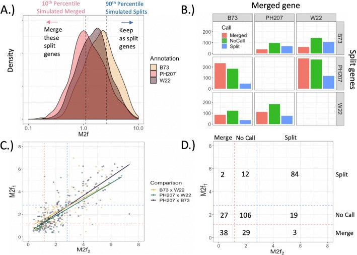
Results of M2f classification. a Observed M2f distribution across all split-genes detected in each annotation. The dotted lines are the threshold values generated by simulating null distributions in Fig. 2c-d. b Number of split-gene candidates (Multiple genes) classified as to whether the split-genes should be annotated as distinct genes or a single, merged gene for each pairwise comparison of annotations. c Correlation of M2f values for instances where a single gene from one annotation corresponded to split-gene candidates in both of the alternative annotations (‘Corroborated’ Merged genes in Fig. 1d). E.g. Each point in the ‘B73 x W22’ comparison corresponds to a single PH207 gene. X-axis is the M2f value from the B73 split-gene candidate, and y-axis is the M2f value from the W22 split-gene candidate. Dotted lines indicate the M2f threshold values in part a. d Joint distribution of classifications across comparisons in part c

