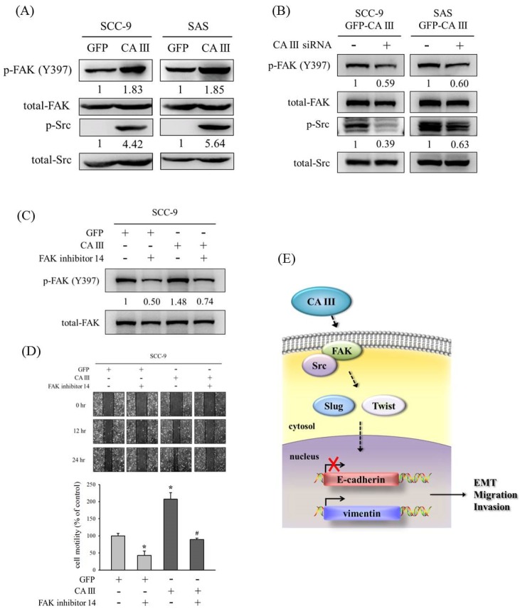
Figure 4.
CA III promotes the migration ability via the FAK/Src pathway in oral cancer cells. (A) The protein expressions of p-FAK (Y397) and p-Src were analyzed by Western blot. β-actin was used as the loading control. Total-FAK and total-Src were used as the loading control. (B) The protein expression of p-FAK (Y397) and p-Src after transfection with scrambled siRNA or CA III siRNA in CA III stable cells. Total-FAK and total-Src were used as the loading control. (C) The protein expression of p-FAK (Y397) after treatment of the FAK inhibitor 14 for 24 h. β-actin was used as the loading control. (D) SCC9-CA III stable cells after the treatment of FAK inhibitor 14 for 24 h were wounded for 24 h. Phase-contrast pictures of the wounds at three different locations were taken. * p < 0.05 compared to GFP stable cells with DMSO. # p < 0.05 compared to CA III stable cells with FAK inhibitor 14. (E) Proposed model for how CA III contributes to the EMT, migration, and invasion abilities in oral cancer.

