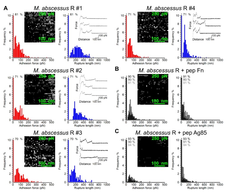
Figure 3.
Single Fn-molecule binding to Ag85 on the surface of M. abscessus R cells. (A) Adhesion force histograms (left) and maps (inset) as well as rupture length histograms (right) and three representative force-distance curves originating from the same dataset for four M. abscessus R cells. (B) Injection of the Fn peptide resulted in significant decreases in Ag85-Fn binding events across the cell surface. Adhesion force histogram plots from three cells were overlaid. (C) Injection of the Ag85 peptide resulted in even greater decreases in Ag85-Fn binding events. In the adhesion maps the z-scale, depicting adhesion force in greyscale, ranges from 0 to 250 pN. Data shown are representative of at least 6 untreated or peptide-treated cells.

