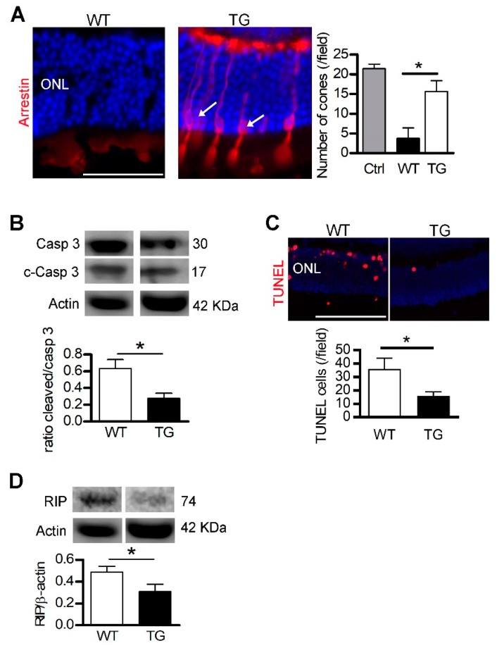
Figure 3.
Transferrin expression preserves the detached retina. Legend Figure 3: After retinal detachment (RD), photoreceptors died by apoptosis and necrosis. Transgenic mice (TG) expressing human transferrin (TF) were used to demonstrate the protective effects of TF. (A) Arrestin staining revealed cones in retinal sections of TG mice (arrows) after RD. Cone number was higher in TG compared with WT mice. (B) The ratio of cleaved/pro–caspase 3 protein level was lower in TG mice compared to WT mice after RD. (C) The number of nuclei positive apoptotic-DNA breaks, stained by TUNEL, was reduced in TG mice compared to WT mice. (D) Necrotic RIP kinase protein level was reduced in TG mice compared with WT mice. All values are represented as the mean ± SEM. Mann–Whitney test (n = 3–6), * p ≤ 0.025. ONL: Outer nuclear layer. Scale bar, 100 µm. From [105]. Reprinted with permission from AAAS.

