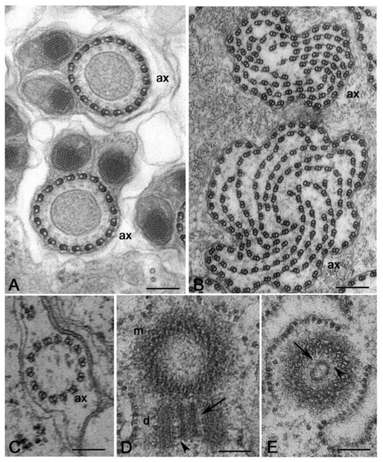
Figure 5.
Axonemes in Hexapoda diverging from the conventional 9+2 model. (A) Cross sections of the axonemes of the spermatids in the cecidomyiids Anaretella showing a circular array of doublets and (B) Monarthropalpus with many doublets arranged in concentric rows. (C) Spermatid of the proturan Acerentomon with an axoneme consisting of 14 doublet microtubules. (D,E) Cross sections of centrioles in the primary spermatocyte of Acerentomon showing that their wall consists of 14 doublet microtubules. The daughter centrioles have an inner large tubule (arrows) connected to the peripheral doublets by thin links (arrowheads). ax, axoneme; d, daughter centriole; m, mother centriole. Bars: 200 nm.

