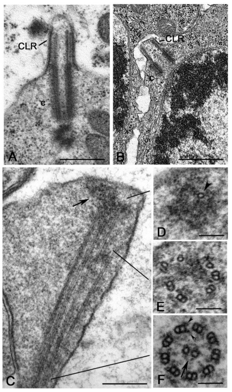
Figure 7.
Ciliary projections in the male germ cells of Hexapoda. (A) Mature Drosophila primary spermatocytes showing full length centrioles (c) projecting out the plasma membrane to assemble a distinct cilium-like region (CLR). (B) Primary spermatocyte of Acerentomon with a short centriole (C) and CLR. (C) Longitudinal section of the distal enlarged region of a ciliary projection in the butterfly Pieris: note the apical cluster of electron-dense material (arrow) in which the tips of some microtubules end. (D,E,F) Cross sections of the ciliary projections showing the gradual organization of the axoneme from the apex to the basal region. (D) Distal sections show a few single tubules (arrowhead) within the electron-dense material. (E) Sections at a lower level display complete and incomplete doublet of microtubules. (F) Proximal sections show complete axonemes consisting of nine doublets of microtubules with dynein arms (arrowheads) and the central tubules (arrow). Bars: A,B, 500 nm; C, 250 nm; D–F, 100 nm.

