Figure 1.
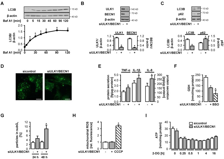
Functional autophagy is a prerequisite for endothelial homeostasis. (A) Human umbilical vein endothelial cells (HUVEC) were treated with 50 nM bafilomycin A1 (Baf A1) for the indicated times. Lysates were subjected to Western blot analyses of LC3B and β-actin. Representative blots and densitometric evaluation are shown (mean values + SEM, n = 5), * p < 0.05 vs. untreated control. (B–I) HUVEC were transfected with control-siRNA or Unc-51-like kinase 1 (ULK1)- plus beclin 1 (BECN1)-siRNA for 72 h and analyzed thereafter (B–F, H–I) or after cultivation for 24 or 48 h (G). (B–C) Cell lysates were analyzed for the indicated proteins in Western blots. Representative blots and densitometric evaluation are shown (mean values + SEM, n = 5). (D) Cells were stained for LC3B, whose accumulation in punctae reflects the formation of autophagosomes. Representative immunofluorescent images are shown (n = 2), scale bar = 10 µm. (E) Cytokines were quantified in cell supernatants by multiplex bead-based flow cytometric analyses (mean values + SEM, n = 5). (F) Glutathione (GSH) levels of cell lysates were determined in a colorimetric assay (mean values + SEM, n = 3). The positive control was treated with 100 µM DL-buthionine-(S,R)-sulfoximine (BSO, inhibitor of GSH synthesis) for 12 h. (G) Cells were stained with propidium iodide and analyzed by flow cytometry. The percentage of particles in the subG1 fraction is shown (mean values + SEM, n = 5). (H) Mitochondrial production of reactive oxygen species was detected by MitoSOX-based flow cytometry. Treatment of cells with 100 µM carbonyl cyanide m-chlorophenyl hydrazone (CCCP) for 1 h served as positive control. (I) Cells were treated with 20 mM 2-deoxyglucose (2-DG) for the indicated times. ATP levels in cell extracts were measured using a luciferase-based assay (mean values + SEM, n = 3). Compared to untreated controls, 2-DG led to a significant reduction of ATP levels under all conditions (p < 0.001, not indicated in the graph). (B–I) * p < 0.05 vs. control-siRNA-treated cells.

