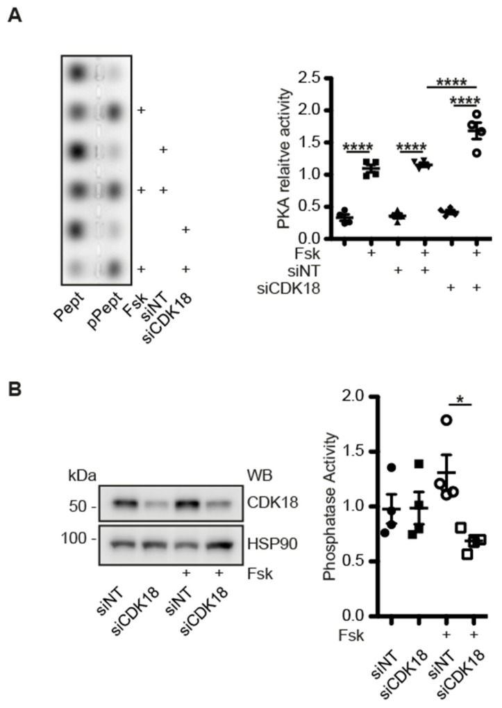
Figure 7.
(A) MCD4 cells were lysed and PKA activity was determined by measuring its ability to phosphorylate a substrate peptide (PepTag A1). Shown is a representative agarose gels from n = 4 independent experiments with PKA-phosphorylated (pPept) and non-phosphorylated (Pept) PepTag A1 peptide. The amounts of each peptide were semi-quantitatively analysed and relative PKA activity was expressed as the ratio of phosphorylated to non-phosphorylated peptides. (B) MCD4 cells were transfected with siNT or CDK18 siRNA, and stimulated with forskolin (Fsk). Phosphatase activity was assayed using para-nitrophenyl-phosphate (pNPP; n = 4). Statistically significant differences are indicated, * p < 0.05, **** p < 0.0001.

