Figure 2.
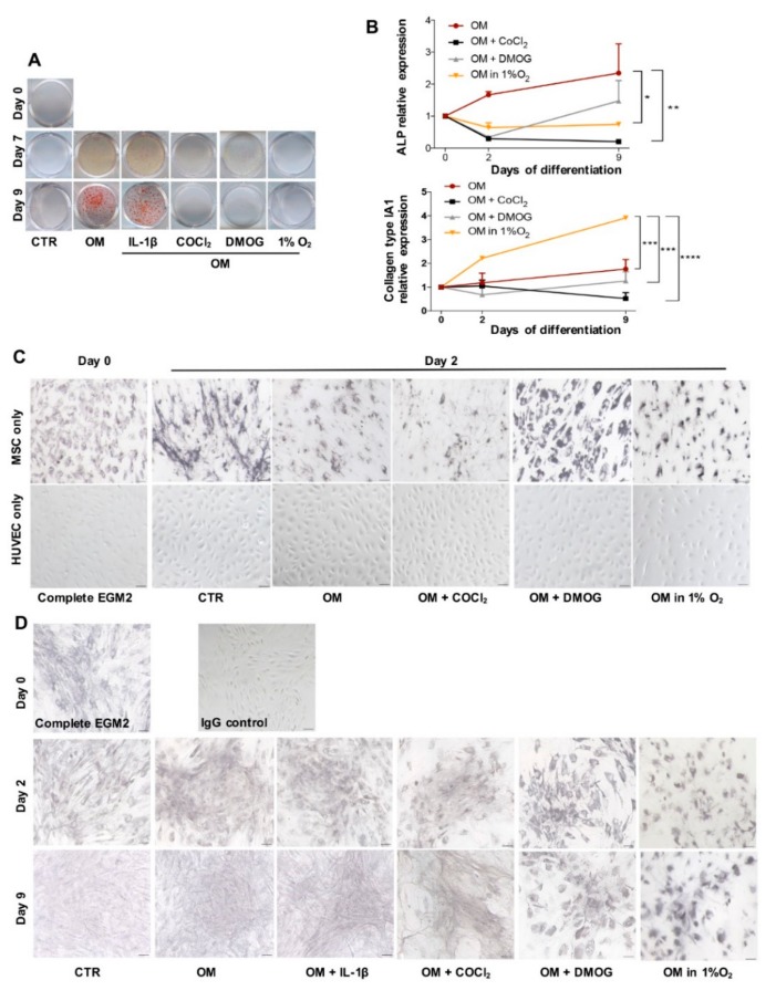
In vitro osteogenesis. (A) Alizarin Red S coloration shows the accumulation of calcium deposit (red) of cells cocultured in control medium (CTR), osteogenic medium (OM), OM added IL-1β or COCl2 or DMOG and OM in 1% O2 at different time points (day 0, 7, 9). (B) qPCR analysis of gene expression of ALP and collagen type I over time (day 0, 2, 9) in conditions OM, OM added COCl2 or DMOG and OM in 1% O2 (n = 3, ∗ p = 0.0156, ∗∗ p < 0.0025, ∗∗∗ p = 0.0002, ∗∗∗∗ p < 0.0001). (C) IHC analysis illustrates Collagen type I expression (black) of BMSC and HUVEC in monoculture in medium Complete EGM2 at day 0 and in day 2 in media CTR, OM, OM supplemented with COCl2 or DMOG and OM in 1% O2. Counterstaining was skipped, scale bar 50 μm. (D) IHC analysis of cocultured cells over time in different conditions. IgG control means the section was not incubated with 1st antibody, scale bar 50 μm.

