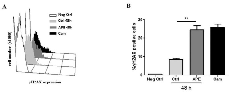
Figure 8.
(A) Distribution diagram of p-γH2AX expression vs. cell number (×1000). (B) Percentage of p-γH2AX positive cells in GB8 cells after 100 μM APE treatment. Camptothecin (5 µM) was used as a positive control. (t-test; APE 48 h vs. Crtl ** p < 0.01). The data are the average ±SEM of three independent experiments.

