Figure 1.
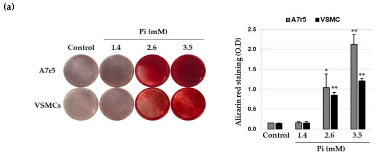
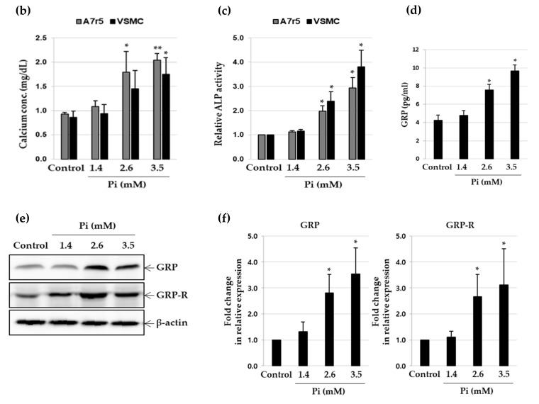
Expression of gastrin-releasing peptide (GRP) and GRP receptor during Pi-induced calcification in vascular smooth muscle cells (VSMCs). A7r5 and primary VSMCs were cultured in calcification medium (1.4, 2.6 and 3.5 mM Pi) for 7 days. (a) Calcium deposition was analyzed by staining with ARS (left), followed by measurement of absorbance to evaluate the degree of mineralization (right). * p < 0.05; ** p < 0.01 vs. control. (b) Calcium content was measured by colorimetric calcium assay. * p < 0.05; ** p < 0.01 vs. control. (c) ALP activity was measured and normalized to protein content, for quantitative analysis. * p < 0.01 vs. control. (d) Secreted GRP content in the conditioned cell culture medium was measured using ELISA. * p < 0.01 vs. control. (e) GRP and GRP receptor (GRP-R) protein levels were examined by western blotting using specific antibodies. β-actin served as the loading control. (f) Total RNA was isolated and analyzed by real-time RT-PCR using the specific primers for rat GRP and GRP-R genes. The expression level of the control (untreated) was set to 1 and the values were normalized to the β-actin mRNA levels. * p < 0.01 vs. control. Data shown are the mean ± SD, obtained from at least three independent experiments.


