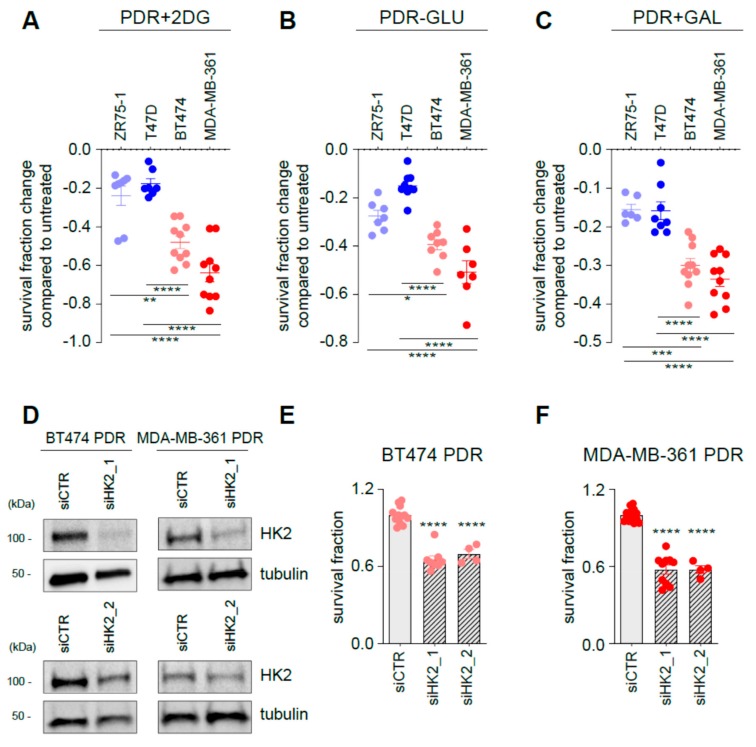
Figure 5.
Targeting glycolysis resensitizes ER+/HER2+ PDR cells to palbociclib. PDR cells were either treated for 3 days in the presence of 2-DG (A), glucose-deprived medium (B) or in a medium in which glucose is replaced by galactose (C). Data are presented as fold change survival fraction of treated versus vehicle-treated or basal-cultured cells. Each dot represents at least three technical replicates from three biological replicate. Shades of blue dots are for HER2− PDR cells, shades of red dots for HER2+ PDR cells. Data represent means ± SEMs and were compared to vehicle-treated or basal-cultured conditions using one-way ANOVA; Dunnett corrected; * p < 0.05; ** p < 0.01; *** p < 0.001; **** p < 0.0001. (D) Total protein lysates from ER+/HER2+ PDR cells transfected with the oligos as described in the figure for 72 h were subjected to Western blot analysis, as indicated. (E, F) Survival fraction changes were measured in cells transfected as indicated in the Figure. Each dot represents at least three technical replicates from three biological replicates. Data represent means ± SEMs and were compared to non-targeting control siRNA (siCTR)-treated cells using Student t test; **** p < 0.0001.

