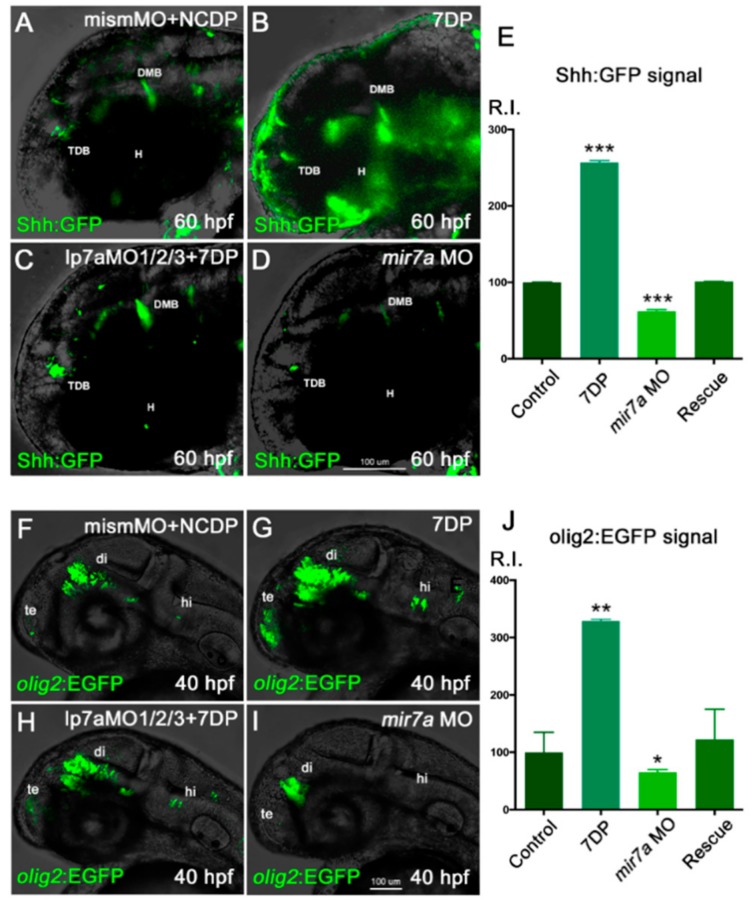
Figure 8.
miR-7 positively regulates Shh signaling responsiveness and olig2+ cells in the zebrafish diencephalon. (A–D) Shh:GFP reporter embryos, injected with NCDP and mismatch morpholino, were used as controls (A). Injection of miR-7 7DP (B) or miR-7a morpholino alone (D) elicits opposite effects on Shh-responsive cells located in the diencephalic region, between the telencephalic-diencephalic boundary (TDB), the diencephalic-mesencephalic boundary (DMB), and the hypothalamus (H), compared with injected controls (A). Co-injection of 7DP and miR-7a MO rescues both conditions (C). All panels show the head region of 60 hpf embryos in lateral view, with the anterior to the left. (E) Chart refers to A–D experimental series. Error bars represent SEM; the experiments were repeated thrice. Sample size n = 5 measures/condition for quantification using Volocity software; (***) p < 0.001. (F–I) olig2:EGFP embryos, injected with NCDP and mismatch morpholino, were used as controls (F). Telencephalic (te), diencephalic (di) and hindbrain (hi) expression of olig2-dependent EGFP appears increased in embryos overexpressing miR-7 7DP (G), and reduced in miR-7a morphants (I). Co-injection of miR-7a MO and 7DP rescues the phenotype (H). All panels are lateral views of the head region at 40 hpf, with the anterior to the left. (J) Chart refers to F–I experimental series. Error bars represent SEM; the experiments were repeated thrice. Sample size n = 5 measures/condition for quantification using Volocity software; (*) p < 0.05; (**) p < 0.01.

