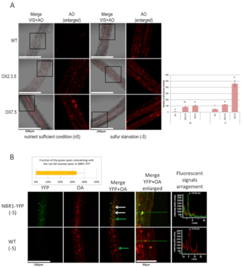
Figure 2.
Comparison of the roots stained with acridine orange (AO). (A) Sulfur starvation increases the number of acidic compartments stained with AO in WT, OX2.3.5, and OX7.5. The graph showing the average number of AO-stained red spots calculated with ImageJ software from three independent experiments (three pictures per each line in each experiment; n = 9) is shown below the representative confocal fluorescence microscopy photographs. The significantly different samples are indicated with different letters (B) NBR1-YFP co-localizes with many acidic compartments stained with AO (examples are shown by arrows: the green arrow points to the dot for which fluorescence signals were arranged in graph view). The results showing the quantification of the colocalization of the green spots with the red spots are from three independent experiments (three pictures each; n = 9).

