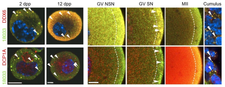
Figure 6.
Germ-cell granules or ‘P-bodies’ in young mouse oocytes and a sub-cortical mRNA storage domain in growing/mature oocytes. Confocal images of mouse oocytes from 2 days postpartum (dpp) and 12 dpp females, immature NSN (non-surrounded nucleolus) and mature SN (surrounded nucleolus) GV oocytes, MII oocytes, and cumulus cells from adult females after staining with 18,033 (stains P-body protein EDC4), DCP1A, and DDX6 antibodies. Diagonal arrowheads depict P-bodies and horizontal arrowheads depict subcortical aggregates. Dashed lines border the subcortical domain. Staining with 18,033 is green, other proteins are red, and DNA staining in blue. Scale bars: 10 μm. Source: Flemr et al., 2010 [28].

