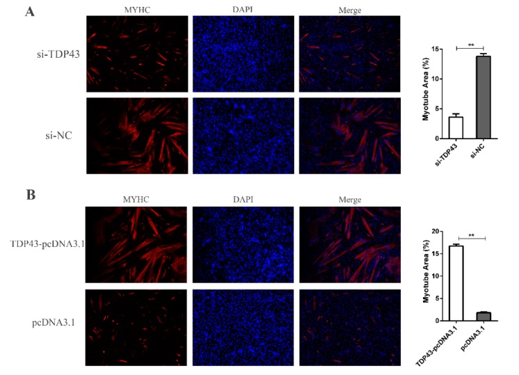
Figure 3.
(A) Representative photographs of MYHC immunofluorescence staining in PSCs differentiated for 24 h showing that knockdown of TDP43 significantly decreased the MYHC expression level. (B) Representative photographs of MYHC immunofluorescence staining in PSCs differentiated for 24 h showing that TDP43-pcDNA3.1 significantly increased the MYHC expression level. Mean values ± SD, n = 3. * means p < 0.05, ** means p < 0.01.

