Figure 1.
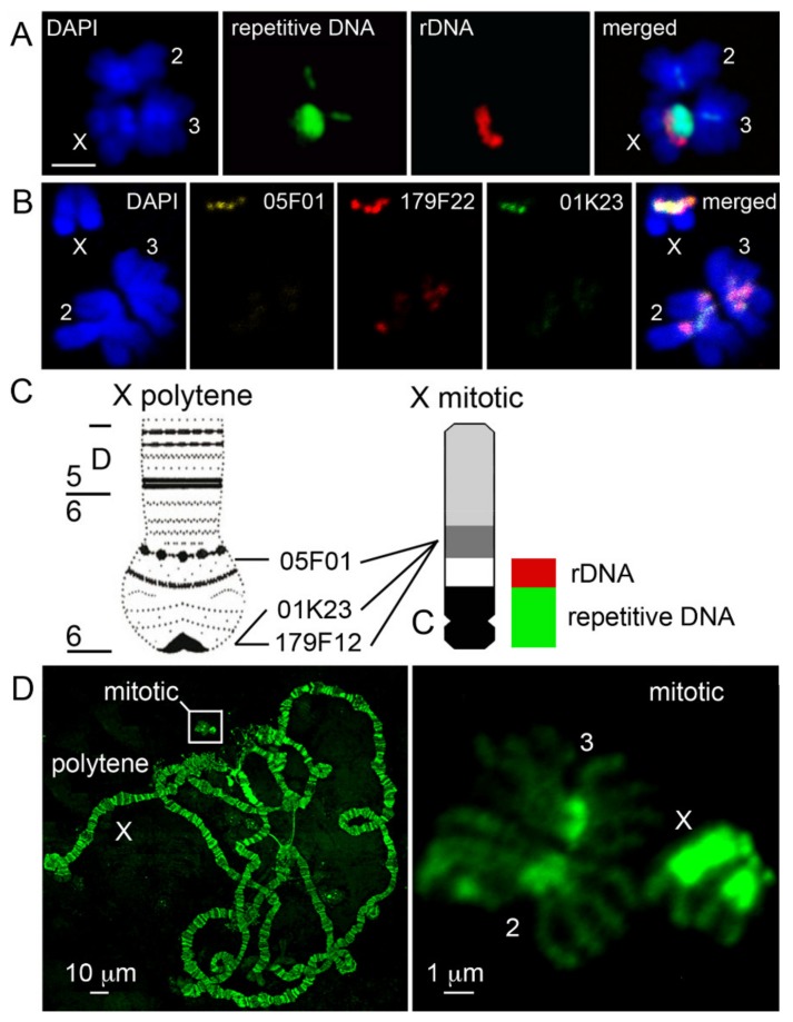
Comparison of the X heterochromatin structure between mitotic and polytene chromosomes. (A) Multicolor fluorescence in situ hybridization (FISH) of a C0t1 fraction of repetitive DNA and 18S rDNA with mitotic chromosomes of An. gambiae PIMPERENA; scale bar is 2 µm. (B) FISH mapping of BAC clones 05F01 (yellow), 179F22 (red), and 01K23 (green) to the distal heterochromatin block on the X chromosome of the An. coluzzii MOPTI strain. Chromosomes are counter-stained with DAPI. (C) Relative positions of heterochromatic blocks and DNA probes of the polytene and mitotic X chromosome idiograms. rDNA = 18S rDNA probe. Repetitive DNA = C0t1 fraction of repetitive DNA. C = putative centromere. (D) Relative sizes of mitotic and polytene chromosomes in the KISUMU strain of An. gambiae. Chromosomes are counter-stained with YOYO-1. Polytene chromosome physical map is from [38].

