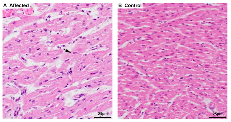
Figure 1.
Histopathology of the heart. (A) Cardiomyocytes of an affected puppy are swollen and pale, and the sarcoplasm around the nucleus is dispersed (arrow) by finely granular material, hematoxylin and eosin (HE) stain. (B) Normal, smaller, and more intense staining of cardiomyocytes of a 5-week-old control puppy, HE stain.

