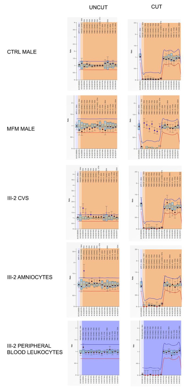
Figure 3.

Graphical representations of MS-MLPA analysis at the FMR1 locus performed on Family 2 proband’s DNA during prenatal and postnatal diagnoses. Only probes of the FMR1 locus are shown. Left panels represent the results before the digestion with methylation-sensitive HhaI enzyme, while the right ones are those after the cut. The HhaI-sensitive probes are indicated in square brackets. In control male (after HhaI cut) the genomic DNA methylation drops to zero due to the absence of FMR1 methylation and thus the enzyme could cut the DNA. On the contrary, in a MFM male, FMR1 probes remained uncut due to the presence of DNA methylation. In proband III-2, FMR1 methylation levels fall after HhaI digestion, because the FMR1 locus is unmethylated as in a normal control male. FMR1 gene was unmethylated both in prenatal (CVS and amniocytes) and in postnatal (peripheral blood leukocytes) samples.
