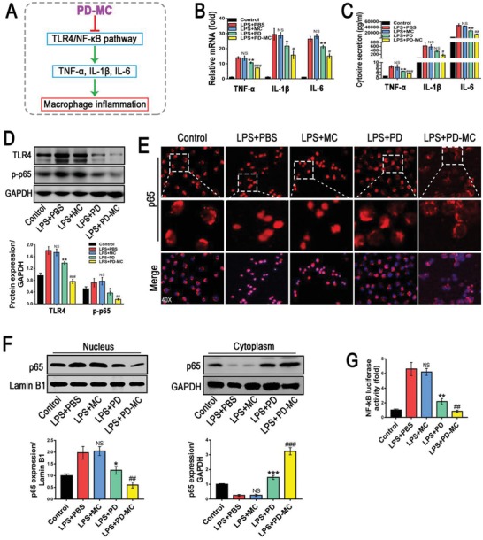
Figure 2.

Anti‐inflammatory efficacy of PD‐MC in RAW cells challenged by LPS. A) Schematic illustration of the anti‐inflammatory mechanism of PD‐MC in macrophages. B,C) The mRNA and secretion levels of TNF‐α, IL‐1β, and IL‐6 were measured by qRT‐PCR and ELISA, respectively. n = 3. D) The protein levels of TLR4/NF‐κB p65 signaling pathway were measured by Western blot assay. n = 3. E) Nuclear translocation of NF‐κB p65 in RAW cells was revealed by immunofluorescence. F) NF‐κB p65 levels in the cytosol and nucleus were assayed by Western blot. n = 3. G) The NF‐κB luciferase activity in RAW cells was measured by luciferase reporter assays. n = 3. NS, no significance; *p < 0.05 and **p < 0.01 versus LX‐2 induced by LPS. #p < 0.05, ##p < 0.01, and ###p < 0.001 versus LPS induced LX2 treated with free polydatin.
