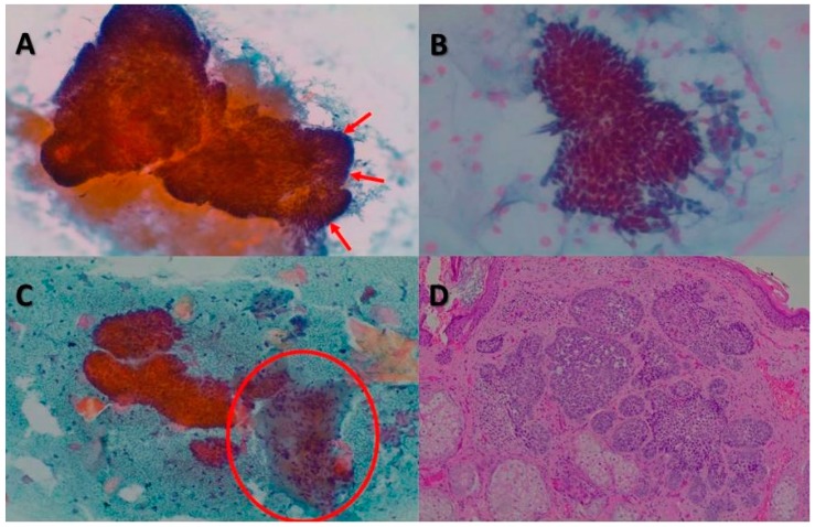
Figure 2.
BCCs. (A) Cytology revealed large clusters of basal cells, mucin, and peripheral cells arranged in palisades (red arrows) (Papanicolaou stain, ×100). (B) Cytological image with a fragment composed of small basal cells with uniform oval, dark nuclei (Papanicolaou stain, ×200). (C) Group of basal cells accompanied by a stromal fragment (red circle) and keratinized squamous cells (Papanicolaou stain, ×100). (D) On histology, BCC showed a lobular pattern with islands and basaloid cells (hematoxylin and eosin, ×40).

