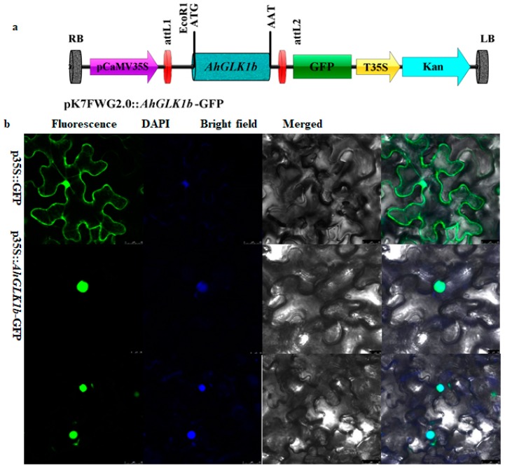
Figure 3.
Nuclear localization of AhGLK1b in tobacco leaves by transient expression using the agroinfiltration method. (a) Schematic diagram of the green fluorescent protein (GFP) vector with the p35S promoter. (b) Localization of AhGLK1b in the nucleus. The fluorescence signal was detected in epidermal cells using a confocal microscope, nucleus was stained with DAPI. The empty-vector pK7FWG2.0 was used as a control. pK7FWG2.0::AhGLK1b-GFP expressing translationally fused AhGLK1b-GFP and cells were visualized at different planes. Scale bar = 638 µm.

