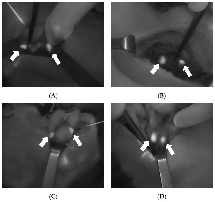
Figure 2.
Four intraoperative images showing the autofluorescence of the parathyroid glands (arrows) as detected with Fluobeam LX® (Fluoptics©, Grenoble, France). (A): Two PGs after right lobectomy. (B): Two PGs after left thyroid lobectomy. (C): Two PGs after superior pole dissection and the medialization of the left thyroid lobe. (D): Two PGs after superior pole dissection and the medialization of the right thyroid lobe. PGs, parathyroid glands.

