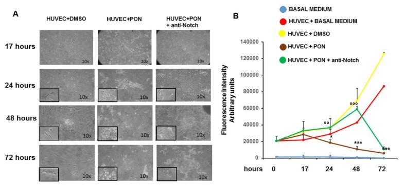
Figure 1.
Effect of ponatinib and Notch-1 signaling inhibition on endothelial proliferation. Human umbilical endothelial cells (HUVECs) were plated at densities of 15,000 per well in 96-well plates and treated with 1.7 nM ponatinib or Dimethyl Sulfoxide (DMSO) for 17 to 72 h, in the presence or absence of 1 μg/mL of neutralizing factor anti-Notch-1 antibody. At the end of each time point, fluorescence intensities were measured with a fluorescence microplate reader using excitation at 485 nm and fluorescence detection at 530 nm. Representative microphotographs showing the effect of ponatinib and Notch-1 signaling inhibition on endothelial proliferation at 10× magnitude, are shown in Panel (A). Insets show the cells at 20× magnitude. Quantitative data (panel (B)) are presented as mean ± standard deviation of fluorescence intensity arbitrary units. n = 3 independent experiments, with at least 7 replicates (culture dishes) per each group. * p < 0.05, *** p < 0.001 ponatinib treated vs. control (DMSO); °° p < 0.01, °°° p < 0.001 ponatinib + anti-Notch-1 antibody treated vs. ponatinib treated. Abbreviations: Pon, ponatinib; DMSO, dimethyl sulfoxide.

