Figure 4.
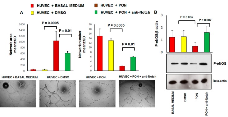
The effect of ponatinib and Notch-1 signaling inhibition on tube formation and endothelial function. Panel (A), Human umbilical endothelial cells (HUVECs) were plated on matrigel-coated plates and treated with 1.7 nM of ponatinib or DMSO, in the presence or absence of 1 μg/mL neutralizing factor anti-Notch-1 antibody. Tube formation was evaluated after 17 h by using light microscopy. Shown are representative fields (5× magnification) of phase-contrast. The pictures are from one representative experiment. Quantitative data (panel (A)) are presented as mean ± standard deviation of several parameters of tube formation. n = 3 independent experiments, with at least 7 replicates per each group. Panel (B), western analysis of phosphorylated eNOS expression in HUVECs treated with 1.7 nM ponatinib or DMSO, in the presence or absence of 1 μg/mL of neutralizing factor anti-Notch-1 antibody, with β-actin serving as a loading control. The bar graph represents for each value the mean ± S.D. from 3 separate experiments. Abbreviations: Pon, ponatinib; DMSO, dimethyl sulfoxide.

