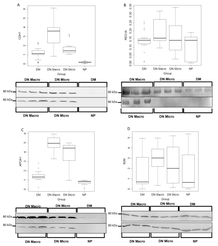
Figure 3.
Western blot Analyses and quantification of the protein excretion in urine of potential marker. Urine samples from 24 patients each group (DM, DN Macro, DN Micro and NP) were collected and proteins were prepared as described in material and methods. CDH1, REG1A, APOA1 and B2M excretion levels in urine were quantified using fluorescence Western blot. Western blot quantification: on the y-axis the line-volume-percentage is given and the x-axis shows distribution of the intensity thought the corresponding urine group where the proteins were analyzed. (A) CDH1, (B) REG1A, (C) APOA1 and (D) B2M

