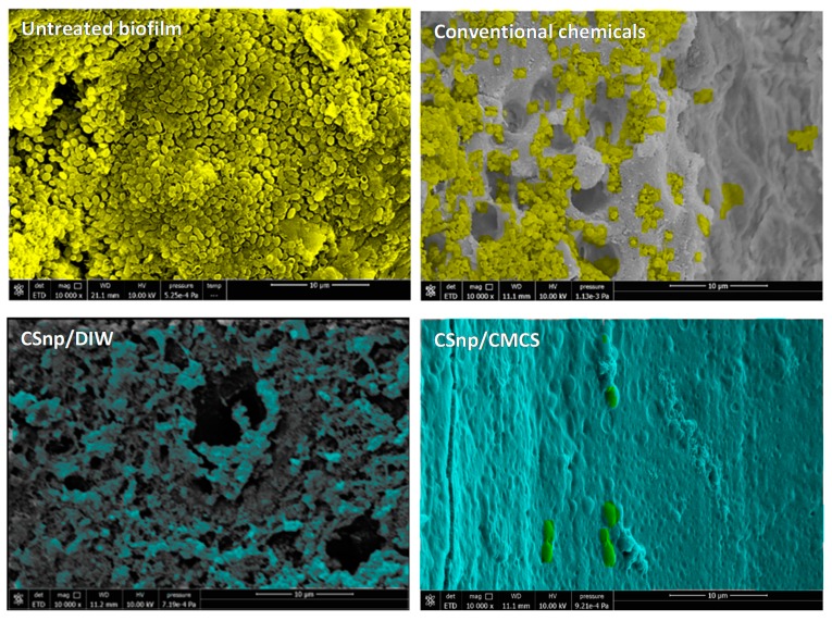
Figure 3.
Scanning electron micrographs showing representative areas of root canal lumen of different groups of 6-week-old E. faecalis biofilm. Untreated biofilm displaying thick bacterial layer on the root canal wall and conventionally treated biofilm showing residual bacteria (pseudo-colored in yellow) versus CSnp treated biofilms which show CSnp-based layer covering the dentin surface (pseudo-colored in turquoise) (10,000× magnification).

