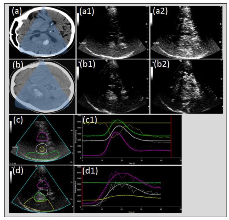
Figure 7.
Perfusion imaging in intracerebral hemorrhage and hemorrhagic transformation of cerebral infarction. Cerebral CT of intracerebral hemorrhage (ICH) of the right basal ganglia, (a) native transcranial gray-scale sonography with hyperechogenic depiction of ICH (a1) and UPI with relative hypo-echogenicity of ICH compared to contrast perfusion of cerebral tissue (a2) due to non-perfusion constricted to the hemorrhagic lesion (c,c1). Cerebral CT of ICH due to hemorrhagic transformation (b), native transcranial gray-scale sonography with hyperechogenic depiction of hemorrhagic transformation (b1) and UPI with persistent hyperechogenicity of the hemorrhagic lesion due to omitted perfusion of the surrounding tissue due to acute stroke (b2) with slowed or missing tissue perfusion (b2,d,d1).

