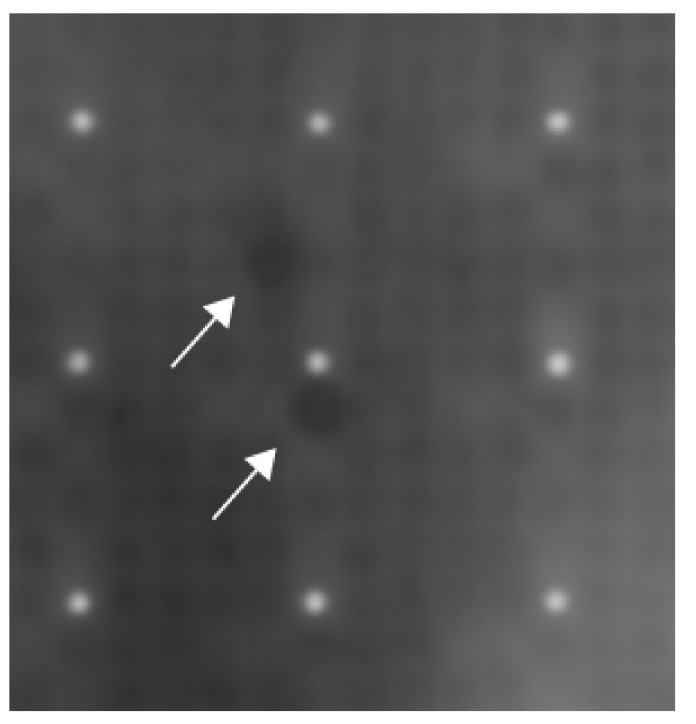
Figure 2.
Exemplary result of one of the three protein-arrays using the sera of patients with sarcoidosis. Each white dot is surrounded by 24 grey dots and together they form a cluster within the protein array. The grey dots contain proteins. Each protein can be found at least two times being apart as far as possible in one cluster. In the center of the picture, two dark spots can be spotted. These two dark spots are Negative Elongation Factor E (white arrows).

