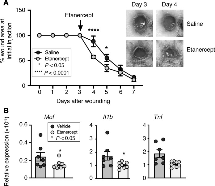
Figure 5. Local TNF-α inhibition improves diabetic wound healing by modulating MOF in wound macrophages.
(A) Representative figure showing improved wound closure after treatment with etanercept. Two wounds were created using 6-mm punch biopsies on the backs of DIO mice. On day 3 after injury, the mice were injected locally with 5 mg/kg etanercept or vehicle control, and change in wound area was analyzed daily using ImageJ software (n = 5 mice/group; repeated twice). Data were analyzed using multiple t tests, 1 per time point. (B) Representative figures showing reduced Mof and inflammatory cytokine expression in response to TNF-α. Wound macrophages (CD11b+CD3–CD19–Ly6G–) were isolated on day 5 after injury from untreated DIO mice and stimulated ex vivo with 250 mg/mL etanercept or vehicle control for 12 hours, and Mof, Il1b, and Tnf expression was measured by qPCR. Data were analyzed using a 1-way Student’s t test with Welch’s correction.

