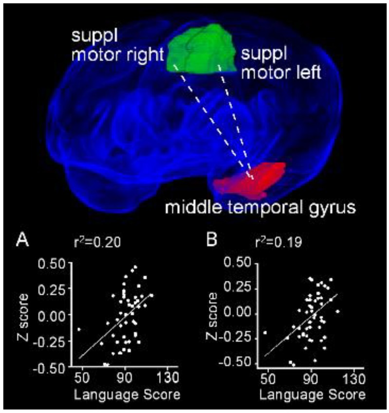
Figure 10.
Resting state functional connectivity between the left middle temporal gyrus and both right and left supplementary motor areas exhibit positive linear relationships with performance on the language subscale of the Bayley III. Obliquely projected 3D reconstruction of the MRI images of cerebral grey matter and identified parcellated regions derived from a neonatal atlas. Dashed white line depicts the connectivity relationship. Inset: Individual Fisher-transformed Z scores of the correlation coefficients between regions. (A) Middle temporal gyrus and right supplemental motor area. (B) Middle temporal gyrus and left supplemental motor area.

