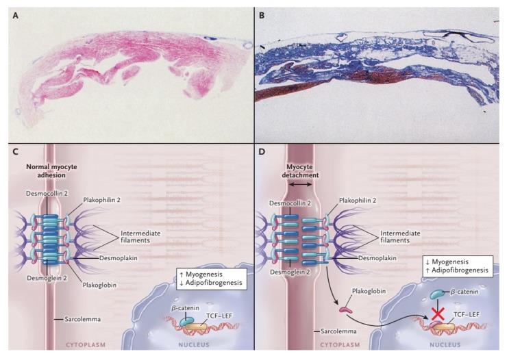
Figure 1.
Histopathological Features and Pathogenesis of arrhythmogenic right ventricular cardiomyopathy (ARVC). With the azan trichrome stain, myocytes appear red, fibrous tissue appears blue, and fatty tissue appears white. Panel A shows a full-thickness histologic section (azan trichrome stain) of the anterior right ventricular wall in a normal heart; Panel B illustrates an analogous section from the heart of a patient with ARVC who died suddenly: Fibro-fatty tissue has replaced the muscular one. Desmosomes are not only structures supplying cell-cell adhesion, but they are also part of the Wnt–β-catenin signaling pathway, which suppresses the expression of adipogenic and fibrogenic genes (Panel C and D). Therefore, on one hand the impairment of desmosomal lead to detachment of cardiomyocytes (double-headed arrow), on the other in a gene transcriptional switch from myogenesis to adipogenesis and fibrogenesis (Panel C and D) [17]. Modified from Ref [4] with permission of the publisher.

