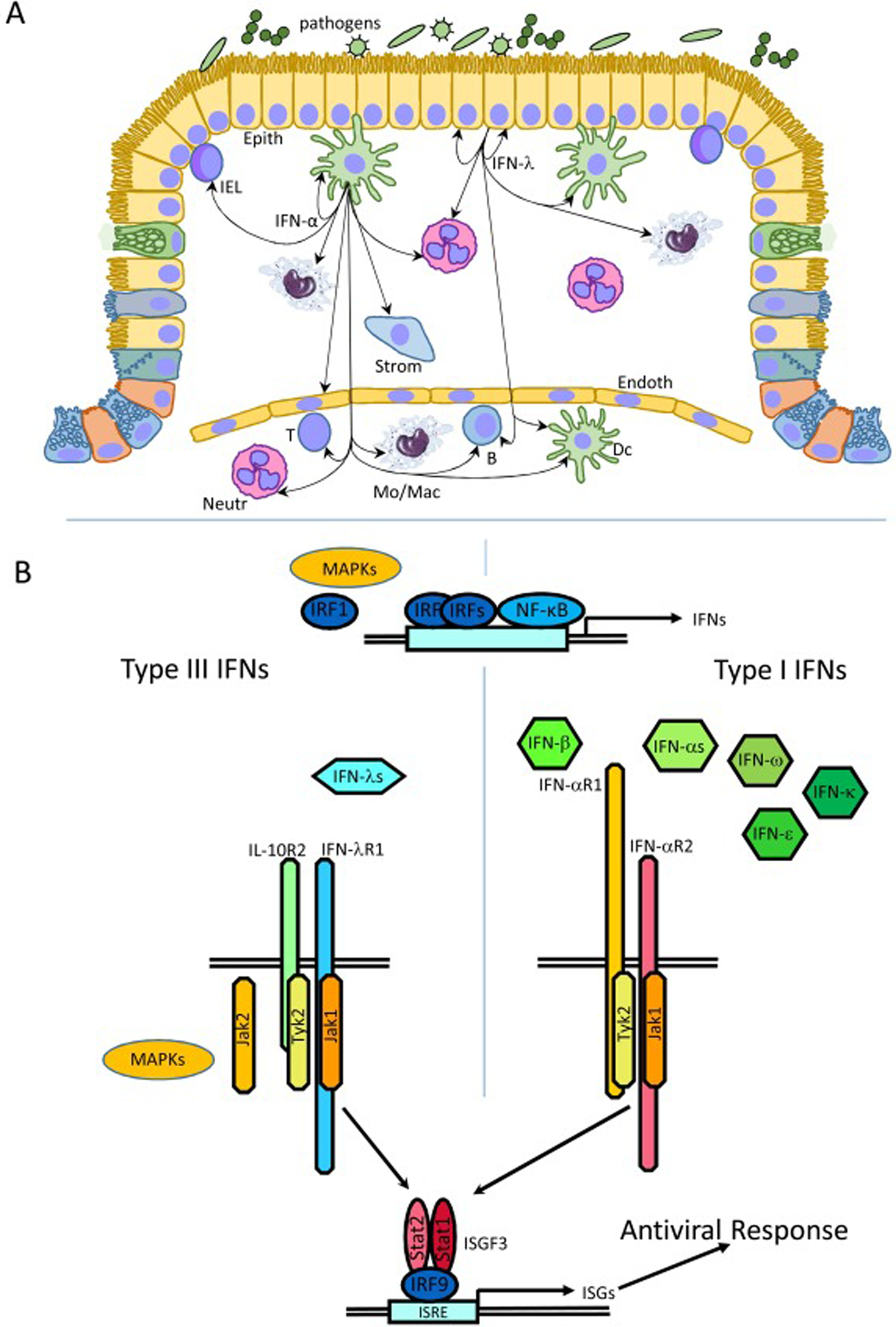
Fig. 1. Division of labor between type I and type III IFNs in antiviral response.

A. Epithelial (Epith) cells are the main source of type III IFNs. Macrophage (Mac), monocytes (Mo) and dendritic cells (DC) can also produce type III IFNs, but primarily produce IFN-αs. IFN-λs act on epithelial cells and tissue-residing neutrophils (Neut), dendritic cells and macrophages, and B cells and plasmacytoid DCs in blood. Most cell types in tissue and in blood including T, B, NK, dendritic, stromal and endothelial cells, Intraepithelial lymphocytes (IEL), macrophages and neutrophils, but not mucosa-lining epithelial cells respond to type I IFNs. B. IRF3, IRF7 and NF-κB are the key transcription factors regulating IFN expression. IRF1 and MAPK signaling has a stronger regulatory effects on type III IFN then on type I IFN production. All type I and all type III IFNs engage distinct IFN-type-specific receptor complexes, but signal through the JAK-STAT pathway resulting in the activation of the same ISGF3 transcription complex and expression of ISGs many of which encode proteins with antiviral functions. Some activities of type III IFNs are also regulated through MAPK signaling and JAK2 involvement.
