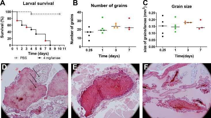
Fig 1. M. mycetomatis infection in G. mellonella larvae over time.
A: Larval survival of PBS infected (---) and M. mycetomatis infected (------) larvae over 10 days, each day is represented with a dot. B: The number of M. mycetomatis grains present in the infected G. mellonella larvae at day 1, 3, and 7 after fungal inoculation as assessed by histology. C: The size of the M. mycetomatis grains present in the infected G. mellonella larvae at day 1, 3, and 7 after fungal inoculation as assessed by histology. D: Hematoxylin Eosin (HE) staining of a M. mycetomatis grain in a G. mellonella larvae, 1 day after fungal inoculation. Arrows, indicate the presence of hemocyte within the cement material of the grain. E: HE staining of a M. mycetomatis grain in a G. mellonella larvae, 3 days after fungal inoculation. F: HE staining of a M. mycetomatis grain in a G. mellonella larvae, 7 days after fungal inoculation.

