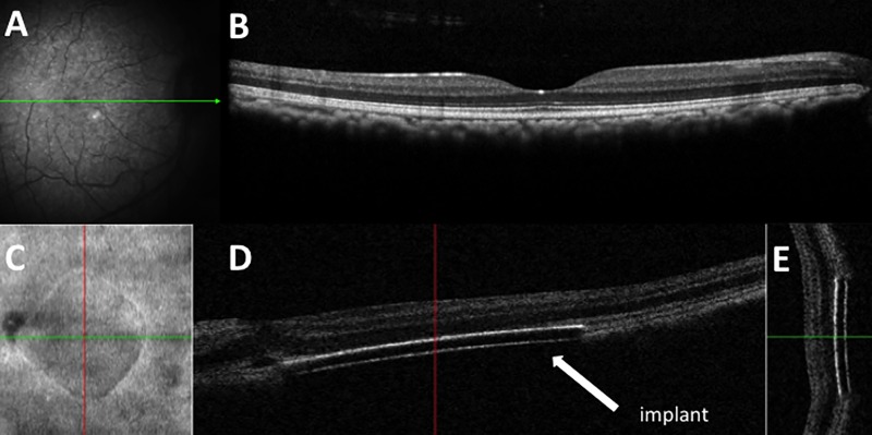
Fig 5. Retinal imaging of a subretinal 2mm PRIMA chip implantation in Primate eye before and immediately after surgery.
A: Preoperative fundus photograph of macula in Macaca fascicularis; B: Preoperative optical coherence scan of macula; C: Enface image of the 2mm retinal implant; D: Horizontal OCT scan showing subretinal location of implant; E: Vertical OCT scan showing subretinal location of implant.

